Scientific Report on
Evolving Methods for Macromolecular Crystallography
Erice, 12 to 22 May 2005
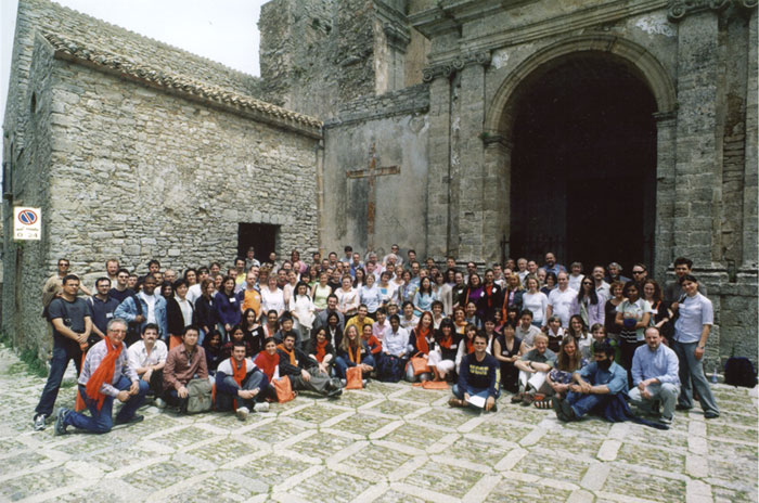
Thanks to thorough planning and hard work by the local organising committee, the school ran smoothly in all respects under their control. The programme was well received by the participants, who informally say that they have all learned a tremendous amount from the oral presentations and hands-on practical sessions.
The major focus of the meeting was on methods used to determine and analyse macromolecular crystal structures. An early highlight was provided by excellent talks on how to obtain useful crystals, by Bergfors and Byrne. Crystals only grow from well-behaved protein, and Terwilliger described how to engineer proteins to adopt a stable, soluble form. Further talks gave clear explanations on how to collect and process the highest quality diffraction data (Garman, Dauter and Leslie). Diffraction measurements alone do not provide the phase information necessary to compute electron density maps. The rapidly-growing collection of known structures can often be used to solve structures by molecular replacement, and two methods to carry this out in difficult cases were presented by Glykos and Read.
Novel structures must be determined by experimental phasing methods based on isomorphous replacement and anomalous dispersion. McCoy and Bricogne presented the theory and practice behind computer programs to obtain phases by these methods. Two talks presented non-traditional approaches, where the anomalous signal from halide soaks (Dauter) or the changes introduced by radiation damage (Ravelli) can be exploited. Once phases are obtained, they can be improved by techniques of density modification (Turk, Emsley and Terwilliger) and in addition by iteratively building progressively more complete models (Perrakis). Once initial models are built they must be refined to agree better with the experimental data (Murshudov). Urzhumtsev described the special insights that can be gained from the small number of protein crystals that diffract beyond atomic resolution, where even the bonding electrons can be observed.
The tremendous progress in recent years in all the techniques of structural biology have made it possible to determine much larger numbers of structures, in what are called high throughput or structural genomics efforts. Computational developments underpinning such efforts were discussed by Adams and Sheldrick. Several structural genomics initiatives were described, including projects aimed at understanding the pathogenic mechanisms of Mycobacterium tuberculosis (Baker) and one aimed at using structure to elucidate biological function (Herzberg). The techniques have matured to the point that it is now possible to determine structures rapidly enough to contribute to responses to emerging pathogens, as Hilgenfeld described for the case of the SARS virus; statistics and controversial measures for this biological danger were widely commented and discussed. .
The consequent structural genomics efforts must be paired with methods to analyse the vast quantity of information contained within three dimensional structures. Patterns in these structures can be detected and then used to identify common enzymatic mechanisms (Thornton). Coupled with data on single nucleotide polymorphisms associated with human disease, they can be used to predict which mutations exert their effects by destabilising the protein (Moult). Some proteins cannot be crystallised at all, or contain large unfolded regions, and the study of large numbers of these now allows unfolded regions to be predicted with reasonable confidence (Sussman). Furthermore, crystal structures of potential drug targets play an essential role in the drug discovery process (Blundell, Turk and van Aalten), and they allow us to understand the molecular basis of some inherited diseases, as Jaskolski showed in the case of amyloid diseases.
The school ended with a look at two developments that push traditional single crystal diffraction studies to new dimensions. Hajdu described how by combining crystallography with spectroscopic studies of the crystal before, during and after data collection, one could be more confident of the exact content of the crystal and could detect radiation-induced chemical changes. Finally, Sayre discussed intriguing new results on diffraction imaging of single particles, showing that a single yeast cell could be imaged in three dimensions to high resolution, using X-ray diffraction.
The selected participants (one per two applicants could join the ASI) were of the highest quality and contributed greatly to the programme, both through oral presentations chosen from submitted abstracts and through well-attended and exciting poster presentations.
In summary, it was a highly successful teaching event that was greatly enjoyed by all participants. The questionnaire item "How do you score (0-100, 100 max.) the value of the course to you?" registered 89 answers with an average of 90.5 (89.9 in 2004)

Randy J. Read
Department of Haematology, University of Cambridge
Cambridge Institute for Medical Research
Wellcome Trust/MRC Building
Hills Road
Cambridge CB2 2XY, U.K.
Tel: + 44 1223 336500
Fax: + 44 1223 336827
E-mail: rjr27@cam.ac.uk
www-structmed.cimr.cam.ac.uk