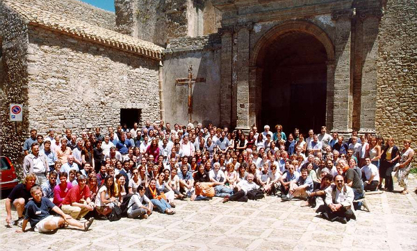
click ON this photo if you wish to see a decent enlargement
Crystallography of Molecular Biology: a Report
E. Majorana Centre, Erice, Trapani, Sicily, Italy: 25 May to 4 June 2000
Since
their inception, crystallographic meetings in Erice have striven to explore
frontier topics.
Additional photos of Erice
and surroundings have been made available by Michael Quayle and Gareth Lewis,
Bristol, participants at Erice 1998.
This
event has been divided into two parallel meetings, both sponsored by the
International Union of Crystallography and by the International Union of
Biochemistry and Molecular Biology, and financed by Nato under the ASI
Programme.
General outline.
The formal lectures were
integrated by several sessions of tutorials and computer demonstrations as organizers
were able to rent or use 20 modern PCs and 7 graphic stations offered for the
meeting by SiG and Compaq. Eighteen selected "students" were given
the occasion to present their work orally and sixtythree could show their
results during poster sessions.
On Sunday 28 the main court of the E. Majorana Centre was named –
during a ceremony – in honour of Dorothy C. Hodgkin,
Nobel Prize 1964 for Chemistry and in the early years Director of the
International School of Crystallography in Erice, who died in 1994. The Erice
Vaciago Prize for the most dynamical young (under 35) "student" in
the lecture hall was awarded to Bostjan Kobe, Slovenian, working in Australia.
A special evening lecture on her May 1999 ten days extraterrestrial flight was
given by the canadian astronaut Julie Payette.
Since the fifth
crystallographic meeting, 1978, an informal questionnaire is handed to all
participants at the end of their stay in Erice ; in June 2000, the question
"Would you like to attend a similar meeting in the future?" resulted in a ratio yes/no 66 to 1 (past
averages 10 to 1 !!! ) ; Erice was
indicated as "the" preferred location 90% of the times. The overall
figure of merit, expressing satisfaction about every
aspect of the meeting, reached the value of 3.18 (maximum 4, minimum 0),
the highest ever recorded.
The Course benefited from important
financial support from Banco di Sicilia, Sede diTrapani and EMBO, and from companies (AstraZeneca, Molndal;
Bristol-Myers Squibb Pharmaceutical Res. Inst., Princeton; Bruker axs, Karlsruhe; Compaq Computer Co., USA;
Cryosystems, Oxford; Hampton
Research, Laguna Niguel, CA; Janssen Pharmaceutica N.V., Beerse;
MAR Research, Norderstedt; Merck
& Co. Inc., USA; Molecular
Structure Co., USA; Novartis,
Basel; Novo Nordisk, Bagsvaerd;
Pfizer, Kent; Pharmacia-Upjohn Research, Nerviano-Milan;
Roche Discovery, Welwyn Garden
City; Schering-Plough Res. Inst., Kenilworth, NJ).
|
|
Methods
for Macromolecular Crystallography |
Supported by the EC, DG
XII, under Contract No. HPCF-CT - 1999 - 00006 : EuroSummerSchools, the first
event of three, short named BIOCRYSTALLOGRAPHY within the series of
CrystalSummerSchools>
Crystallisation is a key
rate limiting step in the analysis of biological macromolecules by X ray diffraction.
Alex McPherson described new approaches to monitoring crystal growth by atomic
force microscopy and DeTitta showed quantitative improvements to automate
crystal growth - 1536 trials simultaneously – for high throughput structure
analysis.
Synchrotron radiation has
revolutionised structural biology. The intense source allows data collection
from very small samples while the tuneable wavelength has allowed a solution to
the phase problem from multi-wavelength anomalous dispersion measurements. Branden
gave an overview of macromolecular beam lines world wide (48 total, 20 in
Europe and 20 in USA) and demonstrated their important advantages for
structural biology and the prospects for the future. Thompson described the
experimental set up at synchrotron source for multi-wavelength measurements
including the parameters of the synchrotron required, the tuneable wavelength,
and the basis of the method for phase determination.
Ealick then took up the
theme of detector development, explaining the physical principles behind
current detectors. Radiation damage by x-ray radiation previously limited data
collection. Garman gave an excellent account of the use of cryomethods (100
°K), that have become so important in the field.
Data processing was
admirably covered by Leslie with lectures and by Otwinowski with hands-on
computer tutorials in small groups: both practical aspects and considerations
about errors - largely appreciated by the younger beginners - that are still
problematic were covered.
Phase determination divided
into ab initio methods (Sheldrick and Hauptman, a special guest, 1985
Nobel Prize for Chemistry) describing their application when high resolution
data (1.2 A) are available and in instances for locating anomalous scatterers
applicable at lower resolution (3A). Only 9% of structures solved in 1999
contained a new fold. Navaza and Rossmann described the powerful methods of
molecular replacement where an existing structure can be used as a starting
point in the interpretation of new structures. This also introduced to the
non-crystallographic symmetry averaging and solvent flattening in phase
determination. Weckert described the 3-beam method for experimental phasing.
Computer graphic displays
and interpretation of electron density maps were covered by Alwyn Jones.
Henderson illustrated efficiently the use of electron microscopy to determine
structures from 2D arrays. These models are finding increasing application as a
starting point for x ray studies of very large structures as exemplified by the
work of Stuart and Rossmann. The convergence of X-ray and EM methods was
finally thoroughly discussed.
There were 113 scientists,
representing 28 nationalities.
|
|
Chemical
Prospectives in Crystallography of Molecular Biology |
This ASI was planned in
order to give to young scientists the "state of the art" of the
Crystallography of Molecular Biology and intended to bring together experimentalists
and theoreticians, biochemists, crystallographers and electron microscopists.
Advances in macromolecular crystallography have given us new eyes to look at
biology. They have led inter alia to an understanding of the rate
enhancement of enzyme catalysed reactions, of control of gene expression, of
regulation by phosphorylation and by allosteric effectors, of recognition in
the component molecules of signal transduction pathways, of recognition in the
proteins of the immune response, of energy transduction, of membrane proteins
and ion channels, protein/nucleic acid complexes, and of viruses, their
assembly from component proteins and their pathogenesis. The most recent
results presented by leader scientists in the field gave to the audience the
idea of the terrific improvements achieved in the understanding. In addition to
the main lectures, young participants had the opportunity to present their
results either as oral presentations or as posters.
E. Getzoff (Scripps, USA)
excited the audience by illustrating how 0.85 Ĺ resolution crystal structures
of the photoactive yellow protein probe the mechanisms of the photocycle on the
nanosecond time-scale. This was followed by a series of lectures by
participants focusing on recent results with signaling molecules. This theme
was further developed by T. Blundell (Cambridge, UK) and J. Kuriyan
(Rockefeller University, USA) who showed information on interacting surfaces in
molecular recognition (e.g. FGF receptors) and molecular dynamics (e.g. Src kinase)
was revealing new biological insights. In the field of immunology, W.
Hendrickson (Columbia, USA) talked on the structural biology of HIV envelope
glycoproteins and their recognition by immune complexes and D.C. Wiley
(Harvard, USA) reviewed how proteins carry out the loading and recognition of
peptides on MHC molecules.
The interactions of
macromolecules and consequences for biological function was also widely
discussed through several examples of structure activity correlations as for
example P. Evans (Cambridge, UK) and A. Brunger’s (Yale, USA) contributions on
the proteins of endocytosis and exocytosis, respectively and S. Harrison
(Harvard, USA) and W. Hol’s (Seattle, USA) talks macromolecular complexes.
Membrane proteins have presented difficult problems in view of difficulties in
their purification and crystallisation. E. Pebay-Peyroula (Grenoble, France)
demonstrated the significant advances made with bacteriorhodopsin crystal
structure and C. Hunte (Frankfurt, Germany) demonstrated the success with the
very large complex of the yeast cytochrome bc1 complex. Protein nucleic acid
interactions are key to understanding the relationship between genes and
proteins. T. Richmond (Zurich, Switzerland) described recent results with the
nucleosome structure, with regard to understanding the roles of the histone
proteins and DNA conformation, and V. Ramakrishnan (Cambridge, UK) described
progress with studies on the 30S subunit of the ribosome, showing how the
ribosomal proteins decorated the extensive RNA core to form a compact
structure.
A session was devoted to
the combined utilization of the Electron Microscopy and X-ray Crystallography
as synergic tools for improving structural knowledge. This brought together
contributions of the experts including A Crowther (Cambridge, UK), K. Downing
(Lawrence Berkeley Laboratory, USA) and W. Kuhlbrandt (Frankfurt, Germany). The
advantages of combination of X-ray and EM was illustrated in Nobel Laureate
Robert Huber’s "EMBO Lecture" on the structures of large protein complexes
for protein degradation and protein folding. The potential of the new third
generation synchrotron facilities was also presented: their extraordinarily
bright beams allow data to be collected from very small crystals. J. Thornton
(London, UK) presented an elegant talk on analysis and validation of protein
structures illustrating the wealth of information available in the protein
structure database and the potential for the future. J Kuriyan (New York, USA)
chaired an evening session arranged as a lively round table discussion on the
contribution by crystallography to structural genomics.
These achievements are
based on many advances in methods and the meeting heard from the experts on the
topics of MAD and SAD phasing (W. Hendrickson & A. Brunger), high
resolution structures (K. Wilson, York, UK), automatic structure interpretation
(T. Terwilliger, Los Alamos, USA and D. Turk, Ljubljana, Slovenia),
non-crystallographic averaging, phase combination and choice of coefficients
for electron density maps (R. Read, Cambridge, UK), maximum likelihood methods
in refinement (E. Dodson, York, UK) and ab initio phasing methods (G.
Bricogne, Cambridge, UK). These sessions were followed by hands on tutorials in
which participants in small groups had the opportunity to learn first hand from
the experts around the computer workstations
There were 114 scientists
representing 27 nationalities..
Lodovico Riva di Sanseverino, email riva@geomin.unibo.it, fax +390 51 209
4904